Showing posts with label MRI. Show all posts

20 February 2014

Working Memory - Studying How The Brain Performs Tasks Based On Memory


Credit: Badre Lab/Brown University
Using Magnetic Resonance Imaging (MRI), researchers at Brown University released a study on how the brain, using working memory, chooses and plans a course of action or task or a series of task.

Similar to short term memory, working memory is a form of memory that keeps and uses information to complete a task or course of action. Working memory is stored in the brain for a limited amount of time; enough time to organize, plan, and execute the task.

The researchers at Brown measured the reaction time of 22 adult volunteers on how the receive, process, and execute a task using working memory. They find that the brain uses an area of the brain called the Caudate and the prefrontal cortex centered on the dorsal anterior premotor cortex. The image above shows the dorsal anterior premotor cortex lit up by MRI.

They also note that working memory uses similar uses similar circuits to those involved in planning motion.

Studying how working memory works helps in understanding brain cognition and also in finding how these parts of the brain affect behavior.

30 August 2013

Time and Brain Coordination Improves Memory Preservation.and Reduces Forgetfulness


Credit: Smithsonian, Photo Researchers, Inc.
Neuroscientist have discovered that coordinated brain activity increases with time after memory formation which results in better memory preservation.

Memory formation in the brain is processed in three stages; encoding, storing, and retrieving. In the encoding stage, information is transformed or encoded by the brain through chemical and electrical signals. Information is then stored in different regions of the brain depending on its type. The retrieval process brings these stored memories into conscious awareness.

The actual process of how memories are formed, stored and then retrieved is still not fully understood.

In this new study, neuroscientists have found that brain coordination during the memory process gets better as time goes by. In their experiment, they noted that a newly formed memory in the test subjects is strengthened when the whole process is reapplied the next day.

10 June 2013

Imaging Individual Molecules Possible Through Magnetic Resonance Imaging and Carbon Nanotubes


Scientists are looking into imaging of individual molecules by using carbon nanotubes and magnetic resonance imaging (MRI). MRI is an imaging technology that uses magnets and radiowaves to construct an internal 3D image of the target.

Graphene is a one atom thick layer of carbon atoms and is considered a 2 dimensional object. The structure of graphene resembles that of chicken wire. When it is rolled up to form a cylinder, it is called a carbon nanotube.

Graphene is one of the strongest material around and is also the thinnest. It also conducts electricity efficiently and is a very good conductor of heat. Graphene is almost completely transparent, yet so dense that even the smallest atom helium cannot pass through it.

Carbon nanotubes also retain these properties and are used in different technologies such as nanotechnology, material science, electronics, and even in bionanotechnology.

27 December 2012

Effective Screening for Alzheimer's Disease Through MRI of the Cerebrospinal Fluid


Using Magnetic Resonance Imaging (MRI) can screen patients for alzheimer's disease effectively. MRI is non-invasive and cost efficient which may be useful to track and monitor the disease as well as observe new treatments for it.

MRI is an imaging application that is usually used in the medical field. It uses magnets and radiowaves to construct an internal 3D image of the target.

MRI has been useful throughout the years in imaging the human body in detail and can also be observed in real time. It tracks the magnetic field of atomic nuclei in the body and is then detected by a scanner that uses it to construct an image.

MRI is also used to effectively image the human brain as well as detect neural activity within it through MRI based imaging techniques such as fMRI (functional MRI) and DTI (Diffusion Tensor Imaging).

The new study on detecting Alzheimer's disease through MRI is an significant step for neurological research. Previous tests for the disease requires a cognitive assessment by the doctor through tests as well as a review of the patient's clinical history and neurological and neuropsychological observations.

20 December 2012

Understanding How The Brain Spatially Represents Object and Action Categories Through fMRI


Scientists uses functional magnetic resonance imaging (fMRI) to study blood and oxygen flow in the brain when it processes and categorizes objects and actions. They are studying how the data is represented spatially in the brain.

Magnetic Resonance Imaging uses the magnetic properties of atomic nuclei to construct a 3 dimensional computer image of the target object. It is a non-intrusive and non-lethal way to image the innermost parts of the body.

Unlike an x-ray where the patient should ingest a trackable isotope for the x-ray to image, the patient in an MRI process just have to lie perfectly still while the MRI's powerful magnets scan the body.

An offshoot to MRI, is functional magnetic resonance imaging or fMRI. With fMRI, doctors track the bloodflow in the patient's brain. This is useful to understand how the brain works and reacts to certain actions, behaviors or stimuli.

fMRI helps scientists understand what part of the brain processes a certain action such as identifying smell, recognizing faces, or even remembering certain memories. Doctors analyze this by identifying what part of the brain has increased blood flow activity during the experiment.

Graphical Visualization of the Group Semantic Space (see Note 1)
Credit: Huth et al., Neuron

25 November 2012

Diffusion Tensor Imaging Show Simple Mental Activities Can Maintain Healthy Brain In Old Age


Researchers collected data using Diffusion Tensor Imaging (DTI) to track brain activity and found a link between regular mental activities such as reading, writing, and playing mental games and healthy aging brains.

Diffusion Tensor Imaging is a type of imaging using Magnetic Resonance Imaging that tracks the movement of water molecules in the tissue or blood vessel. It is useful in studying the structural integrity of a tissue or organ. In the brain study, slow movement of water means that the flow is impeded by structures within the organ. A faster flow would mean that there are less structures in the brain.

There are many natural factors that inhibit the health of the brain when it starts to age. Cell senescence (when the brain cells starts to lose the ability to divide), diet, environment, mental and physical stimulation, and regular brain activity are some of these factors.

17 July 2012

Sodium Buildup in Brain Linked To Multiple Sclerosis Disability and Nerve Degeneration


Multiple sclerosis is an autoimmune disease that affects the brain, optic nerve, and spinal cord. The protective covering that surrounds the nerve cells called Myelin Sheaths are damaged which causes nerve signals to slow down or stop.

The damage is caused when the body's own immune system attacks the nervous system causing inflammation. Diseases where the immune system mistakenly attacks normal body cells or tissues are classified as autoimmune diseases.

Multiple sclerosis (MS) affects women more than men and although the disorder can manifest any age, it is commonly diagnosed between ages 20 and 40.

It is still unknown what causes multiple sclerosis but some factors considered are viruses, family history, or a genetic defect.

There is still no known cure for multiple sclerosis although therapies exist that may slow the progression of the disease. MS treatments focuses on controlling the symptoms to help maintain a normal quality of life.

Sodium buildup in brain linked to disability in multiple sclerosis

A buildup of sodium in the brain detected by magnetic resonance imaging (MRI) may be a biomarker for the degeneration of nerve cells that occurs in patients with multiple sclerosis (MS), according to a new study published online in the journal Radiology.

The study found that patients with early-stage MS showed sodium accumulation in specific brain regions, while patients with more advanced disease showed sodium accumulation throughout the whole brain. Sodium buildup in motor areas of the brain correlated directly to the degree of disability seen in the advanced-stage patients.

"A major challenge with multiple sclerosis is providing patients with a prognosis of disease progression," said Patrick Cozzone, Ph.D., director emeritus of the Center for Magnetic Resonance in Biology and Medicine, a joint unit of National Center for Scientific Research (CNRS) and Aix-Marseille University in Marseille, France. "It's very hard to predict the course of the disease."

In MS, the body's immune system attacks the protective sheath (called myelin) that covers nerve cells, or neurons, in the brain and spinal cord. The scarring affects the neurons' ability to conduct signals, causing neurological and physical disability. The type and severity of MS symptoms, as well as the progression of the disease, vary from one patient to another.

11 July 2012

MRI and Magnetic Particles Track Cells In the Body And Monitor Heart Treatments


MRI stands for magnetic resonance imaging. It allows imaging of the interior of the body without using x-rays or other types of ionizing radiation. An MRI scan is capable of showing fine detail of different tissues.

Magnetic Resonance Imaging (MRI) is an imaging application mostly for medical purposes. MRI uses powerful magnets and radio waves to construct an internal image of the target. It is also known as a Nuclear Magnetic Resonance Imaging (NMRO) or Magnetic Resonance Tomography (MRT).

Unlike an X-Ray where ionizing radiation is bombarded to create an image, a magnetic field is utilized to build and visualize the internal structures in detail.

This is done by having the target or subject lie down within a large powerful magnet. The magnet's magnetic field is used to align the magnetic properties (magnetization) of some atomic nuclei in the body. Radio frequency fields are then used to alter the alignment of this magnetization. The affected nuclei then produces a rotating magnetic field that is detectable by the scanner which then uses the data to form an image.

The magnetic field can be adjusted to construct 2 dimensional or 3 dimensional images. MRI is a handy tool to safely image a human brain since the brain may be sensitive to other imaging techniques. Aside from constructing a brain image, it can also pick up neural activity within the brain.

Tiny magnetic particles may help assess heart treatments

Tiny magnetic particles may help doctors track cells in the body to better determine if treatments work, according to research reported in Circulation: Cardiovascular Imaging, an American Heart Association journal.

Researchers showed that injecting immune cells containing magnetic particles into the bloodstream was safe and did not interfere with cell function. Magnetic resonance imaging (MRI) scans can then track the cells moving through the body.

"This could change how we assess new treatments affecting inflammation and the outcome of a heart attack or heart failure," said Jennifer Richards, M.D., lead author and vascular surgeon at the University of Edinburgh's Centre for Cardiovascular Science in Scotland.

29 June 2012

Mind Reading Device Allow Mute and Disabled To Communicate With Others


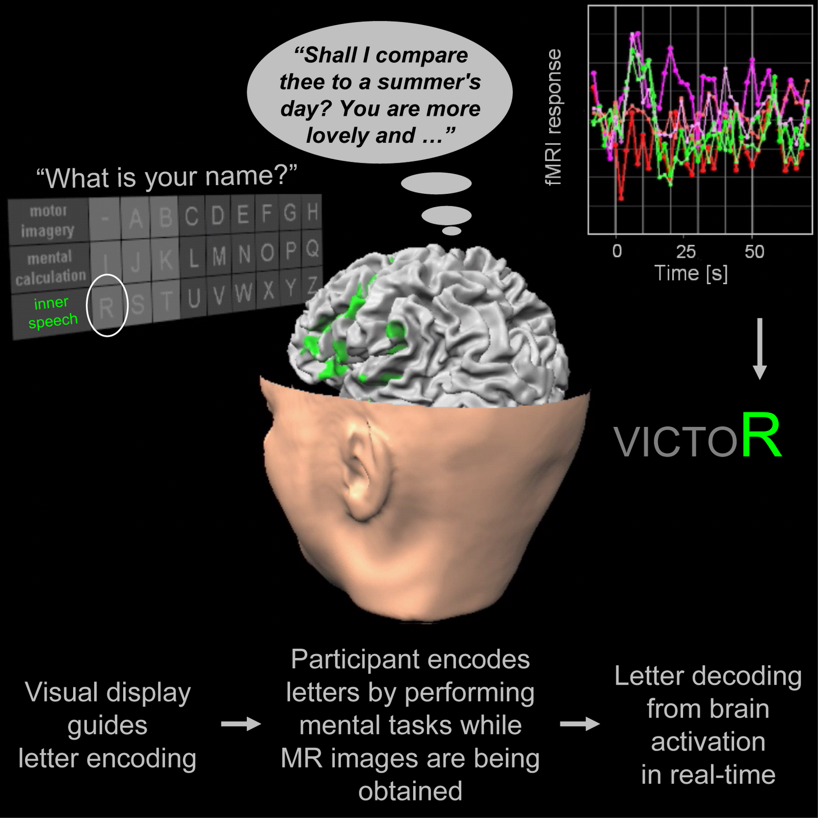
Researchers have come up with a device that may enable people who are completely unable to speak or move at all to nevertheless manage unscripted back-and-forth conversation. The key to such silent and still communication is the first real-time, brain-scanning speller, according to the report published online on June 28 in Current Biology, a Cell Press publication.
Credit: Sorger et al.: Current Biology
Functional Magnetic Resonance Imaging or functional MRI (fMRI) is an imaging procedure using MRI technology that measures brain activity by detecting associated changes in blood flow.

fMRI is used to help diagnose brain diseases and disorders. It also helps doctors study the brain's mental processes to determine how the brain responds to thoughts and feelings.

The technology uses Magnetic Resonance Imaging (MRI). MRI uses radio waves and mgnetic fields are used to create the detailed images of the test subject. In fMRI, imaging is used to study the blood flow in the brain to detect areas of brain activity. A computer records the changes in the blood flow to show the brain was working in real time.

Using fMRI, researchers have developed a device that can help people who are mute or have difficulty speaking to converse with others.

With mind-reading speller, free-for-all conversations that are silent and still

Researchers have come up with a device that may enable people who are completely unable to speak or move at all to nevertheless manage unscripted back-and-forth conversation. The key to such silent and still communication is the first real-time, brain-scanning speller, according to the report published online on June 28 in Current Biology, a Cell Press publication.

The new technology builds on groundbreaking earlier uses of fMRI brain scans to assess consciousness in people described as being in an unconscious, vegetative state and to enable them to answer yes and no questions. fMRI (or functional magnetic resonance imaging) is typically used for clinical and research purposes to track brain activity by measuring blood flow.

"The work of Adrian Owen and colleagues led me to wonder whether it might even become possible to use fMRI, mental tasks, and appropriate experimental designs to freely encode thoughts, letter-by-letter, and therewith enable back-and-forth communication in the absence of motor behavior," said Bettina Sorger of Maastricht University in The Netherlands.

Video: Mind Reading Speller


08 June 2012

Diffusion Tensor Imaging (DTI) With An MRI Gives Insight On How Concussions Differ From Person to Person


Magnetic Resonance Imaging (MRI) is an imaging technique used mostly for medical applications and purposes. It uses powerful magnets and radio waves to construct an image of the body. It is also known as a Nuclear Magnetic Resonance Imaging (NMRO) or Magnetic Resonance Tomography (MRT).

Unlike an X-Ray where ionizing radiation is bombarded to create an image, a magnetic field is utilized to visualize the internal structures in detail.

This is done by having the subject lie down within a large powerful magnet. The magnetic field generated by the magnet is used to align the magnetic properties (magnetization) of some atomic nuclei in the body. Radio frequency fields are then used to alter the alignment of this magnetization. The affected nuclei then produces a rotating magnetic field that is detectable by the scanner which then uses the data to form an image.

The magnetic field can be adjusted to construct 2 dimensional or 3 dimensional images. MRI is a handy tool to safely image a human brain since the brain may be sensitive to other imaging techniques. Aside from constructing a brain image, it can also pick up neural activity within the brain.

Novel brain imaging technique explains why concussions affect people differently

Patients vary widely in their response to concussion, but scientists haven't understood why. Now, using a new technique for analyzing data from brain imaging studies, researchers at Albert Einstein College of Medicine of Yeshiva University and Montefiore Medical Center have found that concussion victims have unique spatial patterns of brain abnormalities that change over time.

The new technique could eventually help in assessing concussion patients, predicting which head injuries are likely to have long-lasting neurological consequences, and evaluating the effectiveness of treatments, according to lead author Michael L. Lipton, M.D., Ph.D., associate director of the Gruss Magnetic Resonance Research Center at Einstein and medical director of magnetic resonance imaging (MRI) services at Montefiore. The findings are published today in the online edition of Brain Imaging and Behavior.